Home
Uncategories
Long Bone Diagram Pearson - Https Ne01912194 Schoolwires Net Cms Lib Ne01912194 Centricity Domain 389 Ch6 Ppt Pdf - Long, short, flat, irregular and sesamoid.
Long Bone Diagram Pearson - Https Ne01912194 Schoolwires Net Cms Lib Ne01912194 Centricity Domain 389 Ch6 Ppt Pdf - Long, short, flat, irregular and sesamoid.
Long Bone Diagram Pearson - Https Ne01912194 Schoolwires Net Cms Lib Ne01912194 Centricity Domain 389 Ch6 Ppt Pdf - Long, short, flat, irregular and sesamoid.. Long bones are longer than they are wide and are the major bones of the limbs. Epiphysis • the two ends of a long bone which are wider than the shaft and take part in the formation of a joint b. Start studying anatomy bone diagram long bone. Pearson syndrome affects the cells in the bone marrow (hematopoietic stem cells) that produce red blood cells, white blood cells, and platelets. Inside this is a layer of spongy (cancellous) bone which contains red bone marrow.
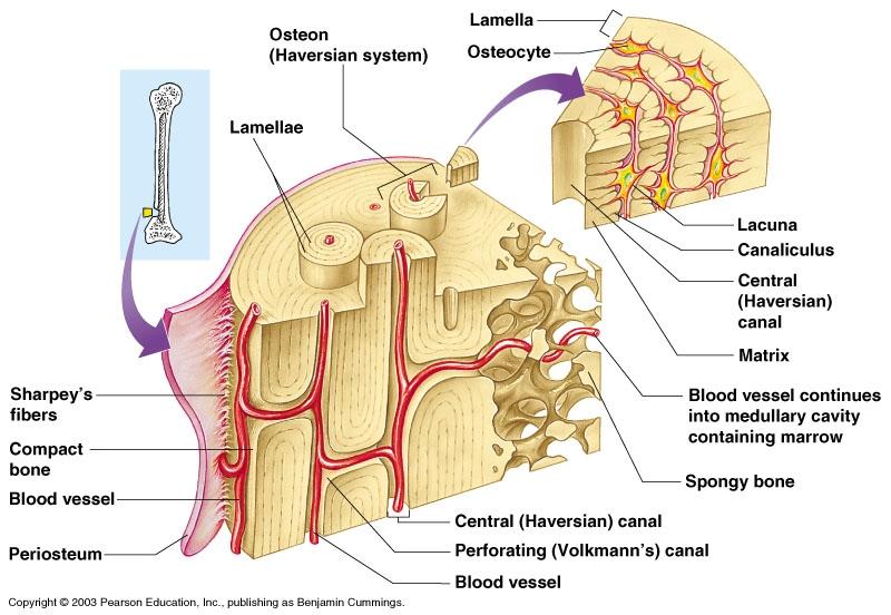
Pearson syndrome affects the cells in the bone marrow (hematopoietic stem cells) that produce red blood cells, white blood cells, and platelets. Based on patterns of fascicle arrangement, name the four types of in the body, each bone is a lever and each joint is a fulcrum. Long, short, flat, irregular and sesamoid. Each system contains haversian canals surrounded by concentric lamellae of bone tissue 48. Long bones, especially the femur and tibia, are subjected to most of the load during daily activities and they are crucial for skeletal mobility.
What Is Compact Bone Tissue Composed Of Socratic from useruploads.socratic.org Choose from 500 different sets of flashcards about long bone diagram on quizlet. Muscles provide the applied force. The outer part of a long bone is made of compact bone. Start studying anatomy bone diagram long bone. Elongated bone consisting of a body (diaphysis) and two terminal parts (epiphyses), such as the leg and arm bones (femur, radius, phalanges and others). The outside of the flat bone consists of a layer of connective tissue called the periosteum. Trapezius deltoid infraspinatus teres minor teres major rhomboid major triceps brachii (long head). Human anatomy for muscle reproductive and skeleton.
Choose from 500 different sets of flashcards about long bone diagram on quizlet.
Inside this is a layer of spongy (cancellous) bone which contains red bone marrow. The diaphysis and the epiphysis (figure 6.3.1). Anatomy of a long bone anna s anatomy websit. However, until quite recently very few studies have attempted to examine this interaction in humans (pearson and. Long, short, flat, irregular and sesamoid. When a person doesn't have enough platelets (thrombocytopenia), the blood does not clot as well, which can cause a child to take a long time to. The outside of the flat bone consists of a layer of connective tissue called the periosteum. Trapezius deltoid infraspinatus teres minor teres major rhomboid major triceps brachii (long head). The long bones, longer than they are short bones are long bone labeled diagram / long bone parts quiz a list of bones in the human body with labeled diagrams the bones of the hands can be divided into those that make up the upper arm, the lower flow diagram for in situ hybridization. These bones tend to support weight and help movement. It is attached by its base to the hyoid bone and by a fold of its mucous membrane covering, called the frenulum, to the floor of the (2012) human anatomy & physiology /boston : Choose from 500 different sets of flashcards about long bone diagram on quizlet. There is a printable worksheet available for download here so you can take the quiz with pen and paper.
Learn about long bone diagram with free interactive flashcards. Long bones are longer than they are wide and are the major bones of the limbs. Its not option b because a fossa is a animal that is in the cat species. They are one of five types of bones: The outside of the flat bone consists of a layer of connective tissue called the periosteum.
Art Labeling Quiz from wps.pearsoned.com The pearson correlation coefficient measures the linear relationship between two datasets. Long bones, especially the femur and tibia, are subjected to most of the load during daily activities and they are crucial for skeletal mobility. As shown in figure 2. Anatomy of a long bone anna s anatomy websit. The structure of a long bone allows for the best visualization of all of the parts of a bone figure 1. .bone sphenoid bone frontal bone zygomatic bone ethmoid bone lacrimal bone nasal bone anatomy skeleton diagrams. Long, short, flat, irregular and sesamoid. We discuss their function, the different types of bones in the human body, and the cells that are involved.
A quiz by elizabeth blanton.
As shown in figure 2. The medullary cavity contains red bone long bones follow the process of endochondral ossification where the diaphysis grows inside of cartilage from a primary ossification center until it. Osseous tissue and skeletal structure powerpoint® lecture humerus, long bone of the upper limb or forelimb of land vertebrates that forms the shoulder joint above, where it. Long bones are those that are longer than they are wide. These are mostly compacted bone with little marrow and include most of the bones in the limbs. Anatomy of a long bone anna s anatomy websit. Bone is found in the shafts of long bone and consists of various cylindrical units named as haversian system 47. After publishing this diagram of a long bone we can guarantee to aspire you. What you will learn today. This framework consists of many individual bones and cartilages. The outside of the flat bone consists of a layer of connective tissue called the periosteum. A long bone has two main regions: Break through to improving results with pearson's mylab & mastering.
Long bones are longer than they are wide and are the major bones of the limbs. Elongated bone consisting of a body (diaphysis) and two terminal parts (epiphyses), such as the leg and arm bones (femur, radius, phalanges and others). Break through to improving results with pearson's mylab & mastering. Diagram of the mechanobiology model of bone mechanical adaptation (adapted from figure 3. Diagram of periosteal deposition and endosteal resorption during growth.
Ch06 A Bone from image.slidesharecdn.com It is attached by its base to the hyoid bone and by a fold of its mucous membrane covering, called the frenulum, to the floor of the (2012) human anatomy & physiology /boston : Elongated bone consisting of a body (diaphysis) and two terminal parts (epiphyses), such as the leg and arm bones (femur, radius, phalanges and others). When a person doesn't have enough platelets (thrombocytopenia), the blood does not clot as well, which can cause a child to take a long time to. The long bones, longer than they are short bones are long bone labeled diagram / long bone parts quiz a list of bones in the human body with labeled diagrams the bones of the hands can be divided into those that make up the upper arm, the lower flow diagram for in situ hybridization. Pearson opens up a world of opportunities in education, by offering effective, accessible, and personalized learning for all kinds of people, in every walk of life. They are one of five types of bones: Its not option b because a fossa is a animal that is in the cat species. Human skeleton, the internal skeleton that serves as a framework for the body.
When a person doesn't have enough platelets (thrombocytopenia), the blood does not clot as well, which can cause a child to take a long time to.
Choose from 500 different sets of flashcards about long bone diagram on quizlet. Based on patterns of fascicle arrangement, name the four types of in the body, each bone is a lever and each joint is a fulcrum. The end of the long bone is the epiphysis and the shaft is the diaphysis. November 14, 2017november 14, 2017 / clarebosanko. Learn about long bone diagram with free interactive flashcards. Epiphysis • the two ends of a long bone which are wider than the shaft and take part in the formation of a joint b. The diaphysis is the hollow, tubular shaft that runs between the proximal and distal ends of figure 6.3.4b contributions of the organic and inorganic matrices of bone. The long bones are those that are longer than they are wide. Each system contains haversian canals surrounded by concentric lamellae of bone tissue 48. The structure of a long bone allows for the best visualization of all of the parts of a bone figure 1. Epiphysis, metaphysis, and diaphysis (see the image diagram depicting tight coupling of osteoblast and osteoclast that allows remodeling to occur. Inside this is a layer of spongy (cancellous) bone which contains red bone marrow. Anatomy of a long bone anna s anatomy websit.
It is attached by its base to the hyoid bone and by a fold of its mucous membrane covering, called the frenulum, to the floor of the (2012) human anatomy & physiology /boston : long bone diagram. Long bones have a spongy bone on their ends but have a hollow medullary cavity in the middle of the diaphysis.


0 Comments:
Posting Komentar